3d bone atlas database
You can rotate models to any angle and zoom in and out Easy to navigate and explore human body. 3D atlas of the bone marrow - in single cell resolution Stem cells located in the bone marrow generate and control the production of blood and immune cells.

3d Vector Reconstruction Of The Muscles Of The Ventral Region Of The Neck From Anatomical Sections Of Korean Visible Human At The Paris Descartes Anatomy Laboratory
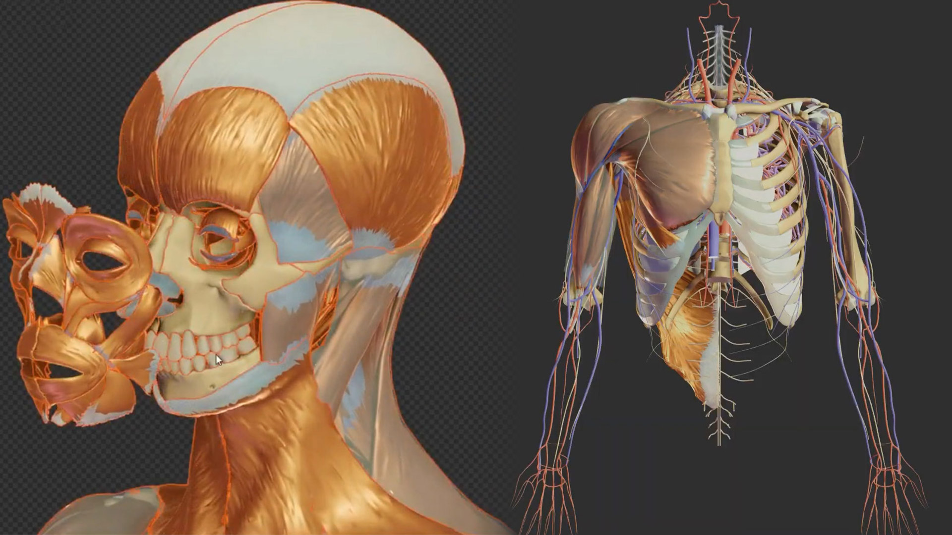
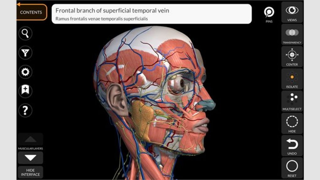
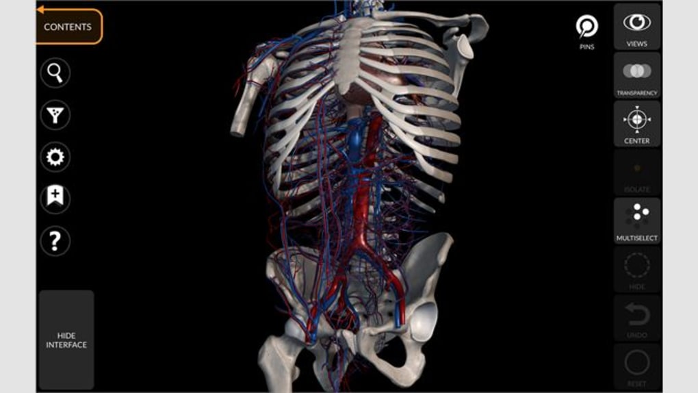
ANATOMY 3D ATLAS allows you to study human anatomy in an easy and interactive way.
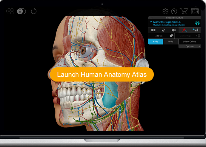
. Intro NOIG Interest Group List What is Oto Is Oto for you Years 1 2 Years 3 4 Research Year No Home Program. The database contains interactive anatomy and physiology learning and visualization content that includes 3D models illustrations and even animations. It has every bone and organ in the human body.
Select parts and Make Embeddable Model of Your Own. 3D Skeletal System. The squamous portion is the largest and smoothest.
3D Temporal Bone Atlas Overview of Anatomy. Comparison of Bone Shapes ツキノワグマ Asiatic black bear 更新情報 2021年12月24日 ツキノワグマを公開しました 2021年11月17日 イヌの手骨格足骨格を公開しまし. Youre born with 33 but the sacrum and coccyx fuse to the rest of the spine making it 24 by the time youre an adult.
BodyParts3D a dictionary-type database for anatomy in which anatomical concepts are represented by 3D structure data that specify corresponding segments of a 3D whole-body model for an adult human male The project was funded by The Integrated Database Project Ministry of Education Culture Sports Science and Technology of Japan. 12 uses a neural network to generate a 3D shape from a single bone radiograph with the goal to recover 3D data for databases of fossils where only 2D data are. From the creator of Visual Anatomy app Features.
3D Temporal Bone Atlas Overview of Anatomy. One of the newest databases that the Weinberg Memorial Library has addedthe Visible Human Body Atlasis an interactive database published by Wolters Kluwer. In 475 of cases 3D model helped redefining the surgical approach Israel Valverde et al.
BONE 3D offers personalized support for the construction of your anatomical model project. On either side of the midline are two rounded elevations. Doesnt matterboth numbers are correct.
The atlas was created by students and embryologists of the Department of. This atlas allows you to scroll through CT slices of the temporal bone in four different planes. - 3 Visualization Modes.
All 3D Models Polygonal only CAD only Free only Sort by. Click on an image to select a plane. 3D Model Reviews no reviews.
Or is it 24. - 3D-HD skull anatomy. Name A-Z Name Z-A Newest Oldest Polys Hi-Lo Polys Lo-Hi Rating Per page.
Highly detailed 3D models with textures up to 4k resolution enable to examine the shape of each structure of the human body with great depth. 3D Atlas Bone 3D CAD Model for AutoCAD SolidWorks Inventor ProEngineer CATIA 3ds Max Maya Cinema 4D Lightwave Softimage Blender and other CAD and 3D modeling software. Atlas Axis and the Atlanto-Axial Relationship Posted on 12612 by Courtney Smith There are 33 vertebrae in your vertebral column.
People Human Anatomy 3D Models Show. General Skull Base and Craniometric Mode. Now the online database which mostly consists of 3D models of art and design pieces has been expanded with a set of human anatomical models with a view to helping out doctors medical students and anyone interested in the human body.
Researchers from EMBL DKFZ and HI-STEM have now jointly developed new methods to reveal the three-dimensional organization of the bone marrow at the single cell level. The temporal bones are divided into the squamosal mastoid tympanic styloid and petrous segments. How To Create 3D Anatomy Photos The 3D photographs featured in this atlas were obtained using an old but enduring technique.
- Anatomical details organised into 10 groups of labels. The squamous orbital and nasal parts. Apart from passive and active 3D viewing platforms like modern televisions and projectors the only way to simulate depth in a 2D environment is by polarizing images for the viewers right and left eyes.
It is completely free. However different organs grow with different. 30 60 90 120 150 180 210 240 270 300.
The frontal bone is made up of three parts. Three-dimensional printed models for surgical planning of complex congenital heart defects. A true and totally 3D free app for learning human anatomy with position quiz built on an advanced interactive 3D touch interface.
Some key figures on the use of anatomical models. We are excited to use Threedings new models says Isaac Cohen an anatomy expert. An anatomy atlas should make your studies simpler not more complicated.
The frontal bone articulates with the right and left parietal bones the zygomatic bones the sphenoid bone the ethmoid bones lacrimal bones maxillary bones and the nasal bones. These skeletal atlases can be displayed from all angles making it easier than ever to understand the characteristics of bone morphology. - 1500 labels and descriptions of.
An international multicentre study. No missing information no confusion and no hidden costs. This database shows the total volume of the embryo grows exponentially with a constant volume increase of 25 per day.
Atlas bone nurbs solid Uploaded by oscar A into People - Human Anatomy 3D Models. We now have created a database of 3D measurement-based skeletal diagrams of the main parts of animal species that are often unearthed from archaeological sites. The approach by Henzler et al.
Osteopathic Applicants FMG Applicants Diversity Inclusion Match 2021-2022. The 3D atlas that contains morphological reconstructions of 14 stages is accompanied by a database with quantitative data based on 17 duplicate stages of reconstructed embryos. The 3D Atlas of Human Embryology comprises 14 user-friendly and interactive 3D-PDFs of all organ systems in real human embryos between stage 7 and 23 15 till 60 days of development and additional stacks of digital images of the original histological sections and annotated digital label files.
Human anatomy simplified with stunning illustrations. Through a simple and intuitive interface it is possible to observe every anatomical structure from any angle. Thats why our free color HD atlas comes with thousands of stunning clearly highlighted and labeled illustrations and diagrams of human anatomy.
3D Bone Atlas Databaseの概要 概要 Overview ヒト Human イヌ Dog イノシシ Wild boar ニホンジカ Japanese sika deer ウシ Cattle ウマ Horse アシカ Sea lion 特集かたちの比較 Special collection. Each articulates with the zygomatic bone zygomaticotemporal suture sphenoid bone sphenosquamosal suture parietal bone parietosquamous suture and occipital bone occipitomastoid suture.

Hand Atlas Of Human Anatomy By Spalteholz Werner 1861 Male Figure Drawing Human Anatomy Anatomy Art

Three Dimensional Images Of Skin Skeleton And Rotated 3d Images Of A Download Scientific Diagram

Axial Skeleton Thoraco Lumbar Spine

3d Anatomical Models Of Eight Organs And The Skeleton Downloaded From Download Scientific Diagram
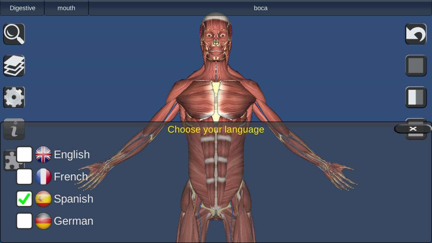
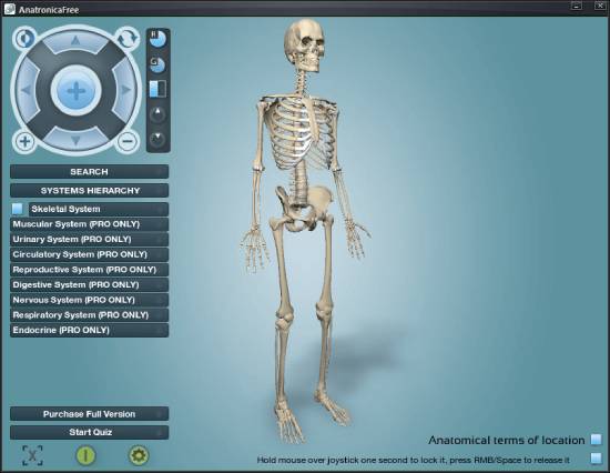
5 Best 3d Anatomy Software For Windows

An Osteometric And 3d Analysis Of The Atlanto Occipital Joint An Initial Screening Method To Exclude Crania And Atlases In Commingled Remains Cappella 2022 American Journal Of Biological Anthropology Wiley Online Library

3d Model Realistic Anatomy Skeleton Muscles
Downloadable 3 D Virtual Models Of The Human Temporal Bone Mass Eye And Ear Otopathology Laboratory
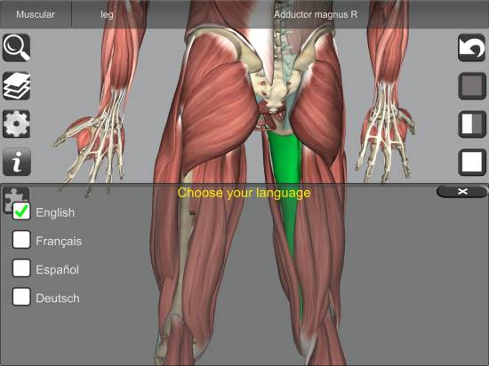
8 Best 3d Anatomy Software For Windows
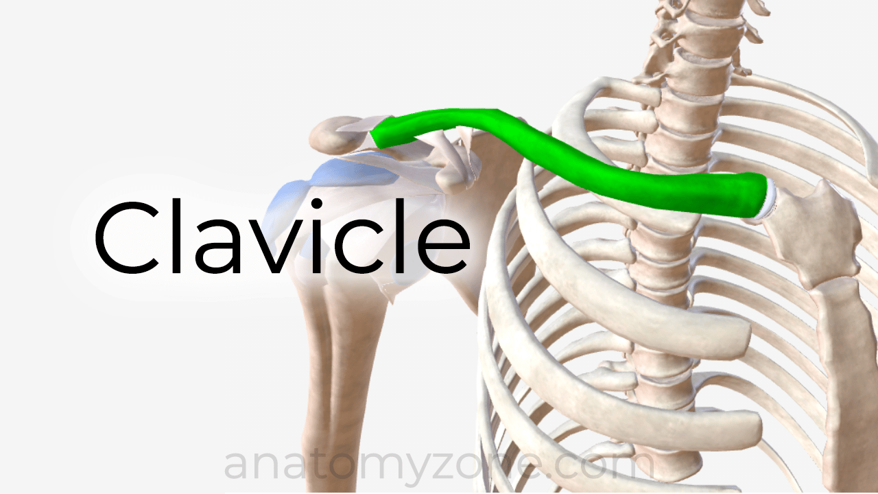
Anatomyzone Your Guide To Human Anatomy

New Visible Body S Human Anatomy Atlas Cmb News Central Medical Library University Of Groningen

Example Of 2d To 3d Registration The Fracture As Seen On The 2d X Rays Download Scientific Diagram
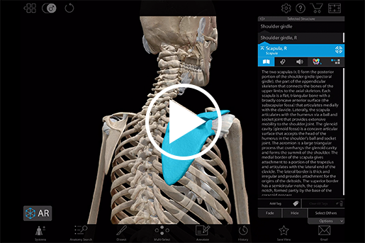
Appendicular Skeleton Learn Skeleton Anatomy
